Most people picture kidney stone surgery as something reserved for big stones. In practice, some of the hardest stones to deal with are small ones. They resist shock wave treatment, or they sit in a part of the kidney that is awkward to reach from below.
In February 2017 I presented my initial experience in Dubai with miniaturised PCNL (keyhole surgery through the back to remove kidney stones), done through a far smaller opening than standard PCNL uses. This post retells that talk.
When a stone needs to come out
Not every kidney stone needs removing. Active removal is considered when a stone is growing, when the patient is at high risk of forming more stones, when the stone blocks the flow of urine or is linked to infection, and when it causes symptoms such as pain or blood in the urine.
Size matters too. Stones larger than 15 mm are usually treated. Smaller stones may be treated when watching and waiting is not the preferred choice, and the decision also weighs the patient's own preference, their other health conditions, and their social situation, such as their work or how much they travel.
One procedure does not fit every stone
Stone treatment has moved a long way since the late 1970s, when a tract (a passage made through the skin into the kidney) was first used to remove stones. Shock wave lithotripsy (ESWL, breaking stones with shock waves from outside the body) later took ground from PCNL. RIRS (retrograde intrarenal surgery, a flexible scope passed up through the urinary passage into the kidney) then took ground from ESWL. Now miniaturised PCNL is taking ground back from ESWL.
My starting point is that every patient needs a tailor-made plan, and no single procedure can be the gold standard for all stones. I look at the site, size and shape of the stone, the anatomy of the kidney, and the patient's other illnesses, and the final decision is taken together with the patient.
Small stones that have not responded to ESWL are a particular challenge. The options for them are RIRS, ultra-mini PCNL and micro PCNL.
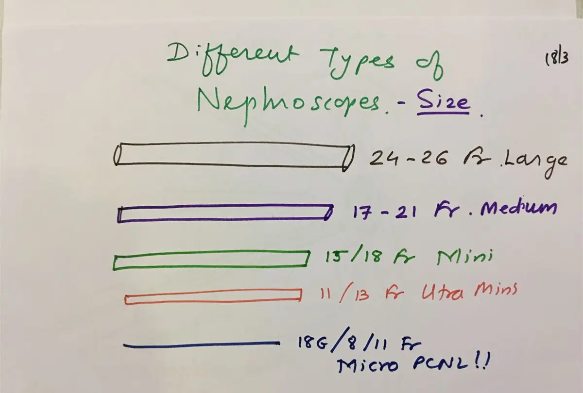
Who I chose it for
In my series I used micro PCNL in selected patients with small kidney stones, from 10 to 18 mm. I did not choose RIRS for them because the angle into the lower part of the kidney was narrow, or the neck of the calyx (one of the cup-shaped chambers that drain urine inside the kidney) holding the stone was narrow.
The stones were in the lower calyces, in calyces with a narrow neck, or in places that could not be reached with a nephroscope (the telescope used inside the kidney) from either direction.
Other situations where the micro system can be used include stones where RIRS access has failed, lower pole stones under 2 cm with difficult anatomy, stones in children, a kidney in an unusual position, as a helper alongside RIRS, as a second access during standard PCNL, and when the patient prefers it.
Planning the entry
The most important step for success, and for avoiding complications, is planning the entry. That means choosing the direction of approach to the stone and understanding the layout of the calyces before the needle goes in.
I used both fluoroscopy (live X-ray) and ultrasound to guide the puncture. The micro system works through what is called the "all seeing needle": a needle 4.85 Fr wide that carries a fine optic, so the surgeon sees the inside of the kidney directly from the moment of puncture. Where the stone burden was larger, I used 8 Fr and 11 Fr sheaths (thin tubes that hold the tract open) instead. A holmium laser was used to break up the stones.
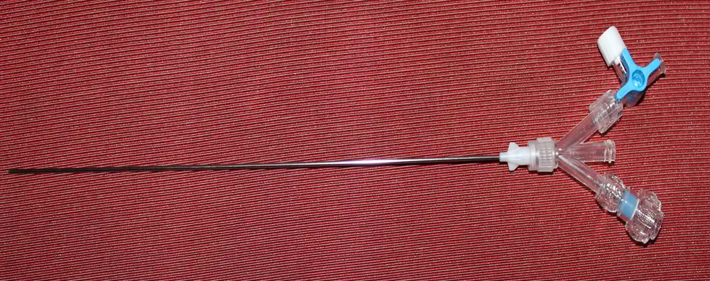
The needle channel takes laser fibres up to 270 microns across. If a larger fibre or another stone-breaking probe is needed, the surgeon changes to an 8 Fr or 10.5 Fr working shaft over a guide wire.
Breaking the stone into dust
Because the channel is so narrow, the stone has to be made very small indeed. Laser settings change what happens. At high energy and low frequency, the laser breaks a stone into fragments: the example I showed was a 2 cm stone broken into 125 pieces of about 4 mm.
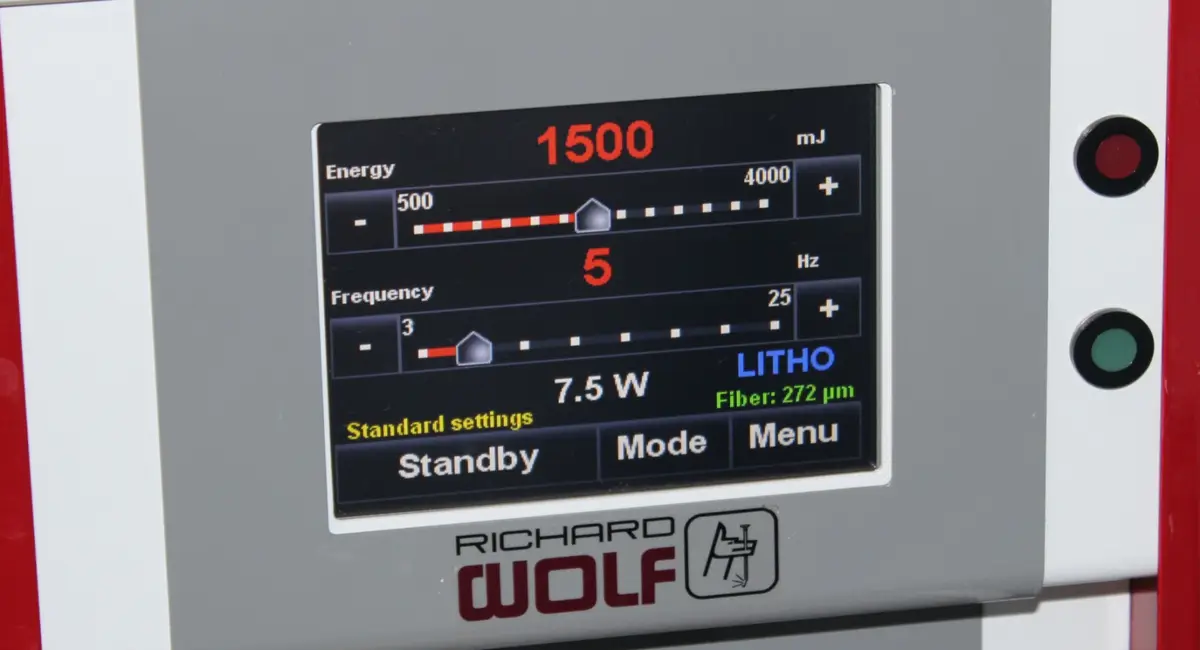
At low energy and high frequency, the laser turns the stone into fine dust. For a large soft stone, I start with low energy and high frequency to create what is called the popcorn effect, and then pulverise the rest into dust.
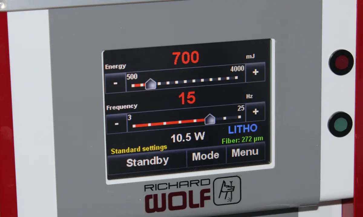
The fragments become small enough to pass down the ureter (the tube from kidney to bladder) on their own. In my series, no nephrostomy (a drainage tube from the kidney out through the back) was placed in 90% of cases, and a feeding tube was placed in the other 10%. Operations done without a nephrostomy are called tubeless; when neither a nephrostomy nor a ureteral stent is left, the procedure is totally tubeless, and in uncomplicated cases this means a shorter hospital stay.
What my first cases showed
I treated 15 renal units (a renal unit is one kidney). Success was judged by ultrasound one month after surgery, with any fragment of 4 mm or less counted as clear. The stone-free rate was 86%. Fragmentation was fast because the laser dusts the stone and the dust passes easily.
The advantages I saw were quick entry, a short recovery, and negligible pain after the operation, with direct sight of the stone throughout. It is still surgery, and the risks remain: urine leak, bleeding, stones left behind, and a rare chance of losing the kidney.
The published studies I showed pointed the same way. In a 2010 study by Cheng and colleagues, blood transfusion was needed in 1.4% of patients after mini PCNL, compared with 10.4% after standard PCNL.
My reasons for valuing this approach were practical. Most stones are smaller than 1.8 cm. Even experts have substantial bleeding at times. Kidney anatomy varies a great deal, and RIRS brings its own complications, may need two or more stages, and raises questions of durability and cost. In a cost-sensitive setting, micro PCNL is the less expensive option.