PCNL (percutaneous nephrolithotomy, keyhole surgery through the back to remove kidney stones) is usually done with the patient lying face down, the prone position. It is the most common approach: well known, easy to learn, and the needle can be guided into the kidney using fluoroscopy (live X-ray imaging).
I prefer to do PCNL with the patient lying on their back, which is called supine PCNL.
What lying face down costs the patient and the team
The prone position carries an anaesthesia risk. Lying on the front is harder for patients who are obese or who have heart and lung problems, and it can press on the vena cava (the large vein that returns blood to the heart).
It also costs time. A thin tube (a ureteric catheter) is first passed up into the kidney from below, with the patient on their back. The patient is then turned over for the PCNL itself. That means two procedures, double scrubbing of staff, double draping, and time spent repositioning.
There is more radiation, stone fragments are retrieved against gravity, and combining the keyhole approach with a telescope passed from below is technically very clumsy.
Why supine PCNL was slow to catch on
The puncture is the most important part of PCNL. The surgeon has to place a needle into exactly the right calyx (one of the cup-shaped chambers that collect urine inside the kidney). If the calyx is missed, the tract may never reach the stone.
In the prone position, X-ray views taken straight on and at 30 degrees let the surgeon judge the puncture in three dimensions. In the supine position, fluoroscopy gives only the straight-on view, so the depth and the point of entry are relatively blind. That is the main reason supine PCNL has been less popular.
Ultrasound solves this, because it gives a three-dimensional entry in the supine position. The calyx can be entered quickly and precisely, with minimal use of radiation.
What the patient and the surgeon gain
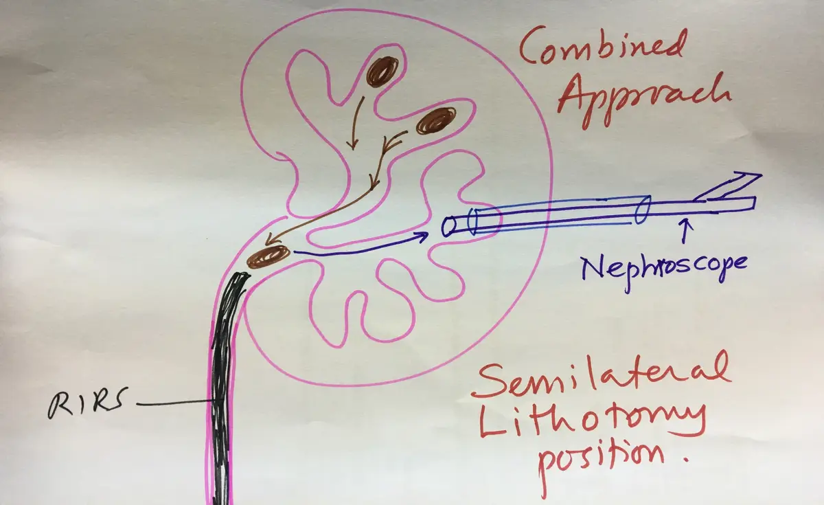
In one position I can place the ureteric catheter, do the PCNL, and, if needed, add a combined approach from below. Total radiation is lower, and so is radiation to the surgeon's hand.
For the anaesthetist, breathing and circulation are undisturbed and the diaphragm moves normally. This suits high anaesthesia risk patients, obese patients, and patients with an unusual body shape.
Because the patient is never turned, there are no shifting injuries and no risk of displacing the breathing tube, drip lines or ureteric catheter. Draping is done once.
In this position the axis of the calyces lies in line with the needle, so the entry is anatomically ideal, and there is less chance of puncturing the colon. Pressure inside the kidney stays low, stone fragments come out by gravity, and the urologist can sit down to operate.
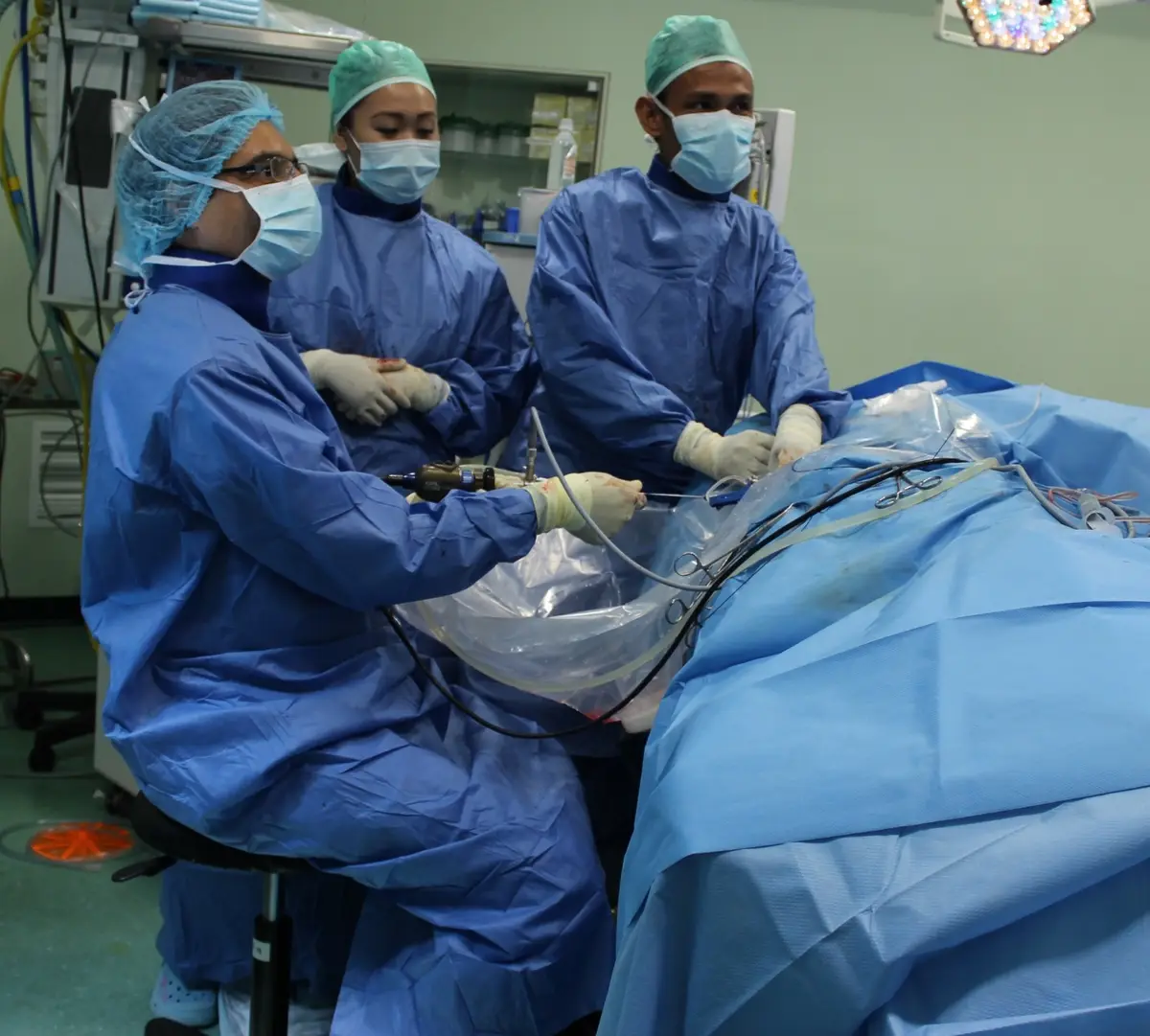
How I plan and perform the operation
Planning starts with the CT scan, reviewed in all three views. I look at the stone, the layout of the calyces, and whether bowel, liver, spleen or pleura (the lining of the lung) lie in the line of the puncture. I choose a line that crosses the least kidney tissue and gives the shortest distance to the stone, and I prefer a dilated calyx.
The aim is a tract that removes as much stone as possible through a relatively straight access. In the supine position the tract is usually through the lower calyx, where the pelvis of the kidney and the upper calyx lie in one line. Stones in the middle calyx may need a flexible scope, passed either through the tract or from below. I also pre-plan the sizes of scope and Amplatz sheath (the tube that holds the tract open).
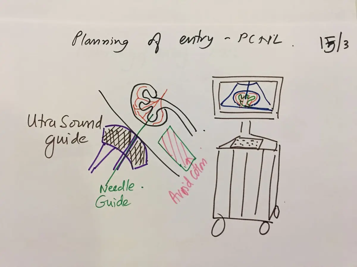
The patient lies on a radiolucent table (one that X-rays pass through), close to its edge, with the legs in supports and the arm on the operated side fixed over the chest. I do a cystoscopy (a look into the bladder with a telescope) and place a 5 or 6 Fr ureteric catheter, then straighten the legs.
I draw landmarks on the skin: the anterior, middle and posterior axillary lines (vertical lines down the side of the chest), the iliac crest (the top of the hip bone), the lower chest line and the ribs. The skin entry is behind the posterior axillary line.
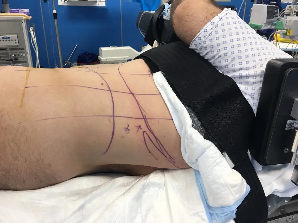
Saline is run up the ureteric catheter to fill the kidney, and I scan it with ultrasound to locate the stone and the preferred calyx. Ultrasound also shows neighbouring organs very easily.
The needle goes through a guide fixed to the ultrasound probe, set at 5 or 25 degrees, and I confirm the end point with fluoroscopy. When the needle hub is removed, saline flows out, which shows the needle is in the collecting system.
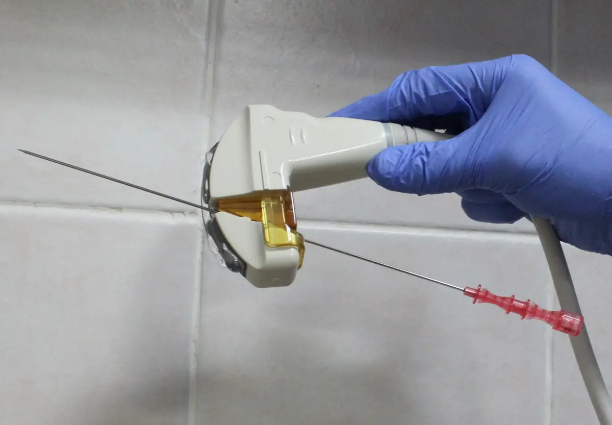
A guide wire goes in, the probe is released, and I make a small skin cut around the needle. The tract is widened with dilators and an Amplatz sheath of 16 to 22 Fr is placed, or a smaller tract is made for ultra-mini or micro PCNL. Stones are broken with a pneumatic or holmium laser device and removed by saline flow or forceps, with the table tilted to help.
The limits of the supine approach, and how I handle them
A difficult puncture calls for a good ultrasound machine with a central needle guide. Upper calyx access is harder, but a supracostal puncture (above the lowest rib) is still possible, and a flexible scope can reach it. Contrast fills the calyces less well, and tilting the head of the table up helps.
The kidney moves more during the puncture, so pressure is applied on the opposite side of the abdomen. Saline distends the kidney less during nephroscopy (looking inside the kidney with a telescope), which I manage with a gauze knot tied around the Amplatz sheath. If the guide wire coils and risks slipping out, RIRS (a flexible scope passed up from below) can catch the wire and pass it down the ureter.
Instruments have less room to move. Keeping the patient at the edge of the table helps, as does a flexible nephroscope, or a combined approach where a scope from below moves stones into a position where they can be removed through the tract.
When I still turn the patient over
In the later version of this talk I listed the cases where I choose the prone position: a horseshoe kidney, a supine puncture line that would cross another organ, a single stone in the upper or middle calyx, or when the supine position does not give an optimal tract.