In November 2018 I taught a hands-on PCNL workshop at the 7th Emirates International Urological Conference. PCNL (percutaneous nephrolithotomy) is keyhole surgery through the back or side to remove kidney stones. The workshop was about how to plan the operation and carry it out safely, and this post follows that thread.
Every patient, stone and kidney is different
Planning starts with three questions. Who is the patient, what is the stone, and what does the kidney's drainage system look like? I weigh the patient's other illnesses, the site, size and shape of the stone, and the anatomy of the kidney.
For complex stones, I rely on a CT IVP (a CT scan taken after contrast dye is injected, so the drainage system lights up). It gives a three-dimensional view of how the drainage system and the stone sit in relation to each other.
Matching the treatment to the stone
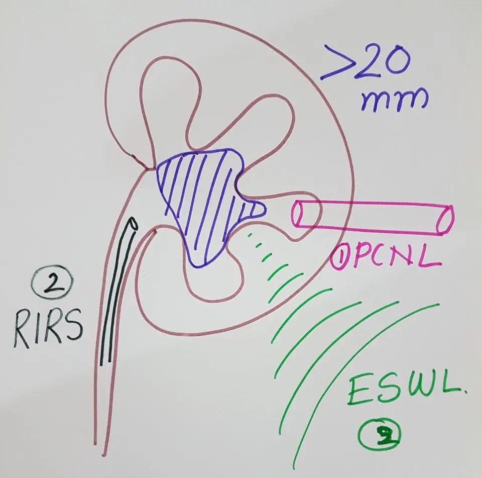
Stone size gives a first order of choice, which I drew out for the workshop. For a stone over 20 mm, PCNL comes first, then RIRS (retrograde intrarenal surgery, a thin flexible scope passed up through the urinary passage into the kidney), then ESWL (shock wave lithotripsy, breaking the stone with shock waves from outside the body).
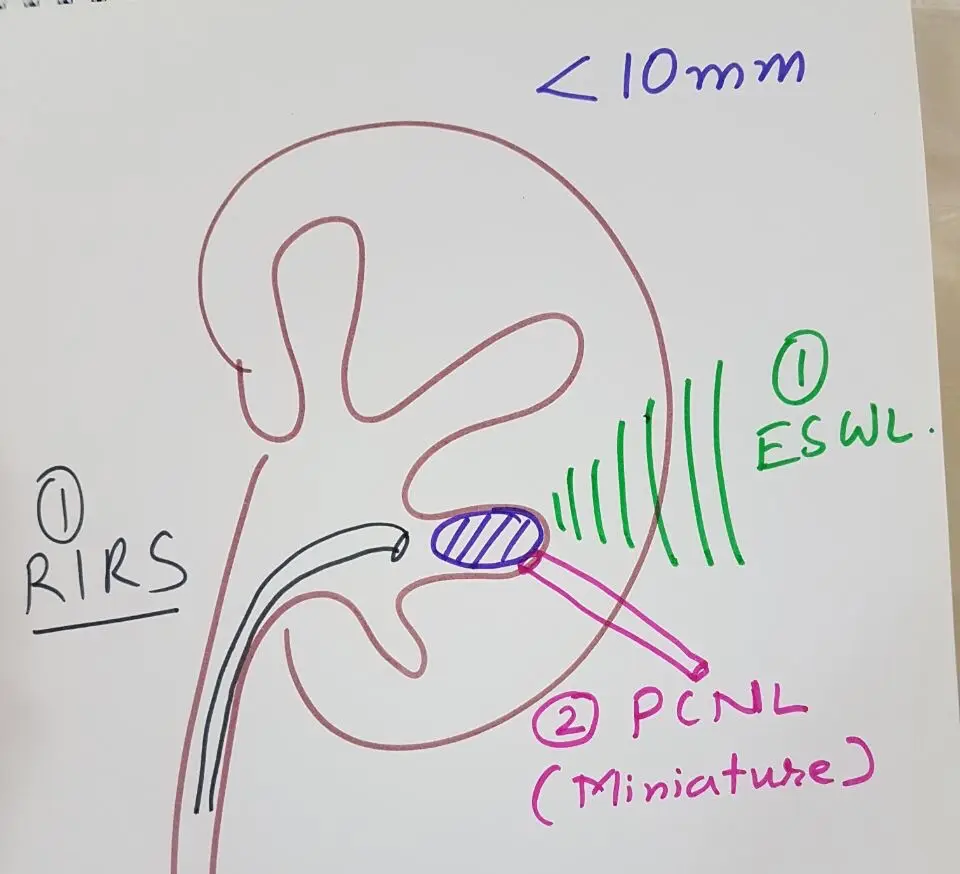
For stones of 10 to 20 mm outside the lower pole, ESWL comes first, followed by endourology (surgery done with scopes), meaning mini PCNL or RIRS. For stones under 10 mm, ESWL or RIRS comes first, with a miniaturised PCNL, such as ultra-mini PCNL or Micro Perc, as the second option.
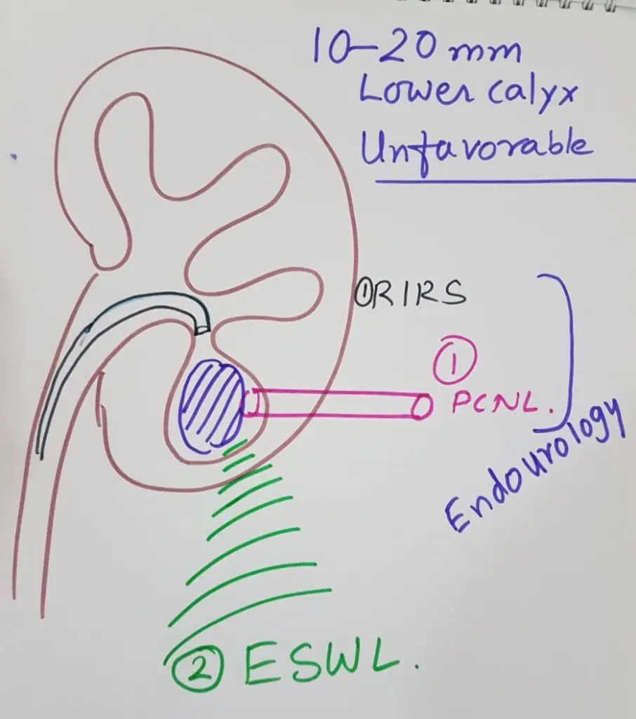
The lower pole of the kidney is a special case. For a 10 to 20 mm lower pole stone with features favourable for ESWL, ESWL still comes first, then RIRS or mini or ultra-mini PCNL. When the features are unfavourable for ESWL, the order reverses: mini or ultra-mini PCNL first, then RIRS, with ESWL last.
Some kidneys need extra thought: a horseshoe kidney (two kidneys joined at the lower end), a kidney sitting low in the pelvis, or a spine curved by kyphoscoliosis.
Planning the entry
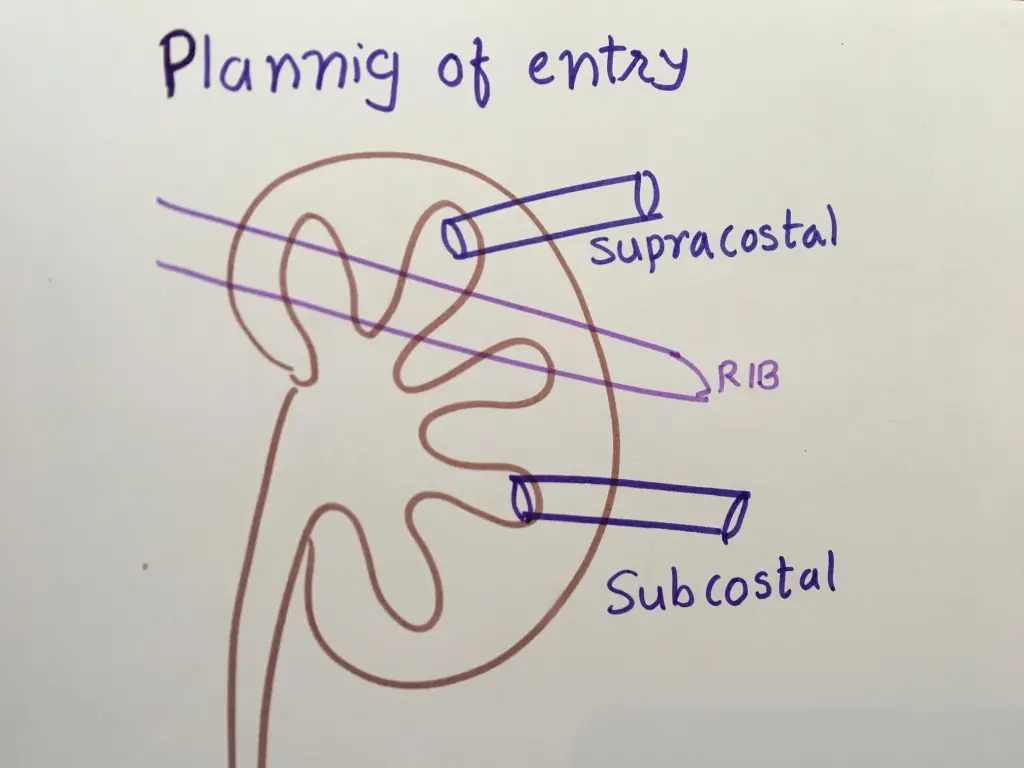
The next step is to decide where the needle goes in. The route into the kidney can pass above the rib (supracostal) or below it (subcostal). Either way, the target is a chosen calyx (one of the cup-shaped chambers that drain urine inside the kidney).
The operation begins with a ureteric catheter passed up from the bladder with a cystoscope. The patient is then turned onto their front. The preferred calyx is located, and the needle is passed into it under fluoroscopy (live X-ray), ultrasound, or both together. After that, a guide wire is placed down the ureter, the tract is widened, and a working sheath is placed.
When breaking the stone, I pointed out one practical difference: the laser does not give a kinetic impact, which lessens the chance of bleeding.
Knowing when to stop
Some decisions are made during the operation. A larger stone left behind is best dealt with through another tract. A stone in the neighbouring calyx can be reached with a flexible nephroscope (the telescope used inside the kidney) or a combined approach. A small leftover stone that the tract cannot reach suits ESWL or RIRS later.
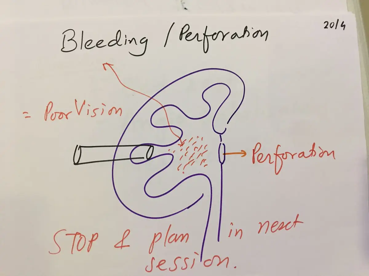
If there is bleeding or a perforation of the drainage system, the right move is to stop. Bleeding makes vision poor, and the next sitting is planned for a few days later.
Working from both ends
For scattered stones with a large total burden, I use a combined approach. The patient lies in a semi-lateral lithotomy position, which allows one surgeon to work up from below and another through the back at the same time.
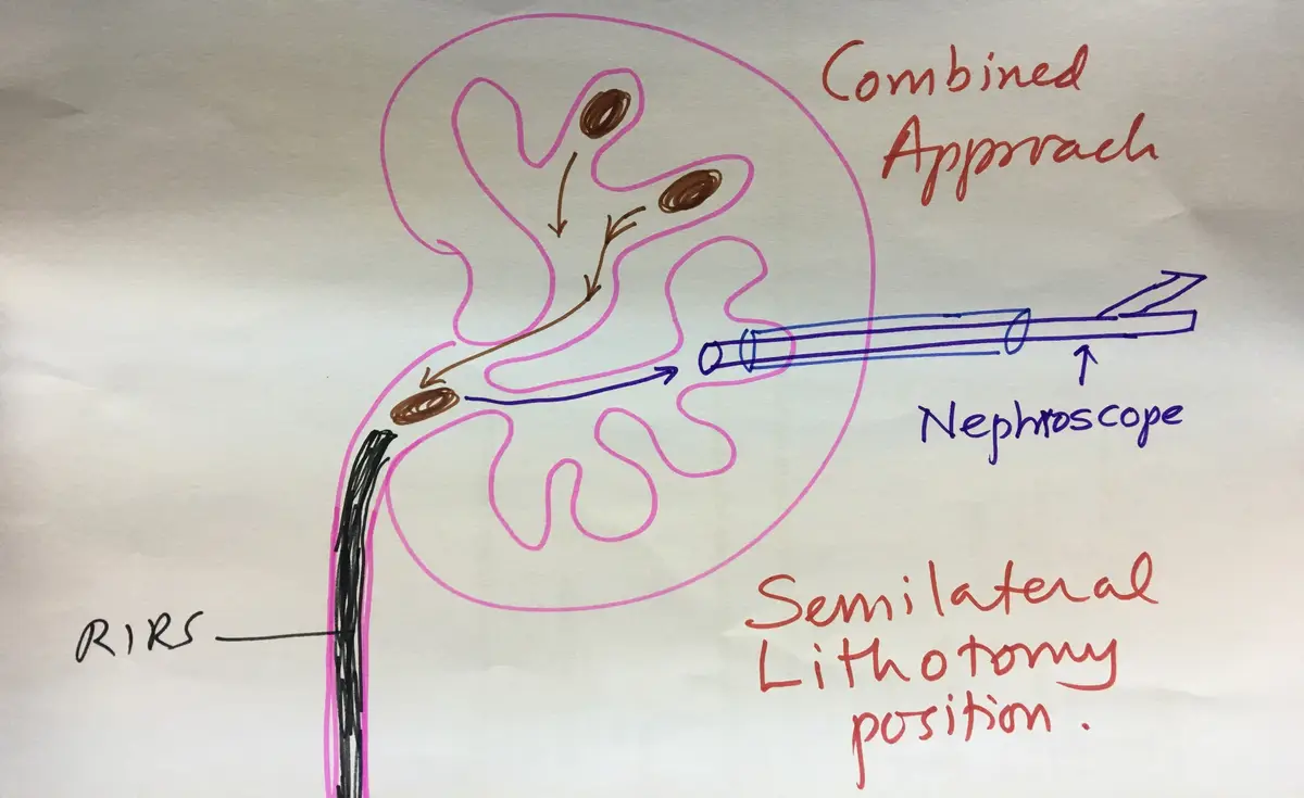
In a fresh case I start with the semi-rigid or flexible scope from below. The stones are broken and moved into the renal pelvis or one calyx. The drainage system stays filled with saline, so manoeuvring is easy, and bleeding from a PCNL tract cannot blur the view. Lastly, a supine PCNL brings the fragments out.
The advantages are that multiple punctures are not needed, stones come out easily with gravity, larger stones are retrieved in less time, and it is faster than RIRS alone. The limitations are two teams and two sets of equipment, which adds cost, and a drainage system that deflates.
Lying on the back instead of the front
Supine PCNL, with the patient on their back, works better with ultrasound guidance. There is no pressure on the vena cava (the large vein returning blood to the heart), the diaphragm moves normally, and it suits obese patients, patients at high anaesthetic risk, and people with an unusual body shape. A scope can be passed from below at the same time.
Pressure inside the kidney stays low and fragments come out by gravity. The urologist can sit down, their hands get less radiation, and no time is lost turning the patient twice. The limitations: it needs a good ultrasound machine with a needle guide, upper calyx puncture is difficult, the calyces fill less with contrast, the kidney moves more during puncture, there is less saline distension, and the scopes have less room to manoeuvre.
Choosing the tract size, and how to finish
Standard access tracts are 24 to 30 Fr (French, the scale used for instrument width). Smaller tracts may bring less bleeding and less pain after surgery, but they mean longer operations and poorer vision from lower saline flow, and small instruments can raise pressure inside the kidney.
At the end of the operation, the options are a DJ stent (an internal tube from kidney to bladder), a nephrostomy (a drain out through the back), or going tubeless. Whether to leave a nephrostomy depends on leftover stones, the chance of a second look, significant blood loss, urine leaking outside the drainage system, blockage of the ureter, infected stones, a single kidney, or a bleeding tendency. When neither tube is left, the operation is called totally tubeless, and in uncomplicated cases it means a shorter hospital stay.
The complications to be ready for are urine leak, bleeding, fever and stones left behind. Bleeding is managed with bed rest, and when needed angioembolisation (blocking the bleeding vessel from inside, through a catheter) or removal of the kidney (nephrectomy).