PCNL (percutaneous nephrolithotomy) is keyhole surgery to remove kidney stones. The surgeon makes a small tract from the skin of the back straight into the kidney, passes a telescope through it, breaks the stone and takes the pieces out. This talk explained what PCNL is, who it suits, and how it is done step by step.
The idea is not new. In the late 1970s surgeons began using a tract through the skin to remove stones, and Arthur Smith pioneered PCNL. Shock wave treatment and flexible telescopes later took over many cases, but PCNL has stayed the standard procedure for large kidney stones.
Which stones need PCNL
A kidney stone needs active removal when it is growing, blocking the kidney, causing infection, or causing symptoms such as pain or blood in the urine. It also applies to patients at high risk of forming stones. The list also includes stones over 15 mm, and smaller stones when simply watching them is not the preferred choice. In the talk I showed these alongside the 2016 European Association of Urology treatment algorithm.
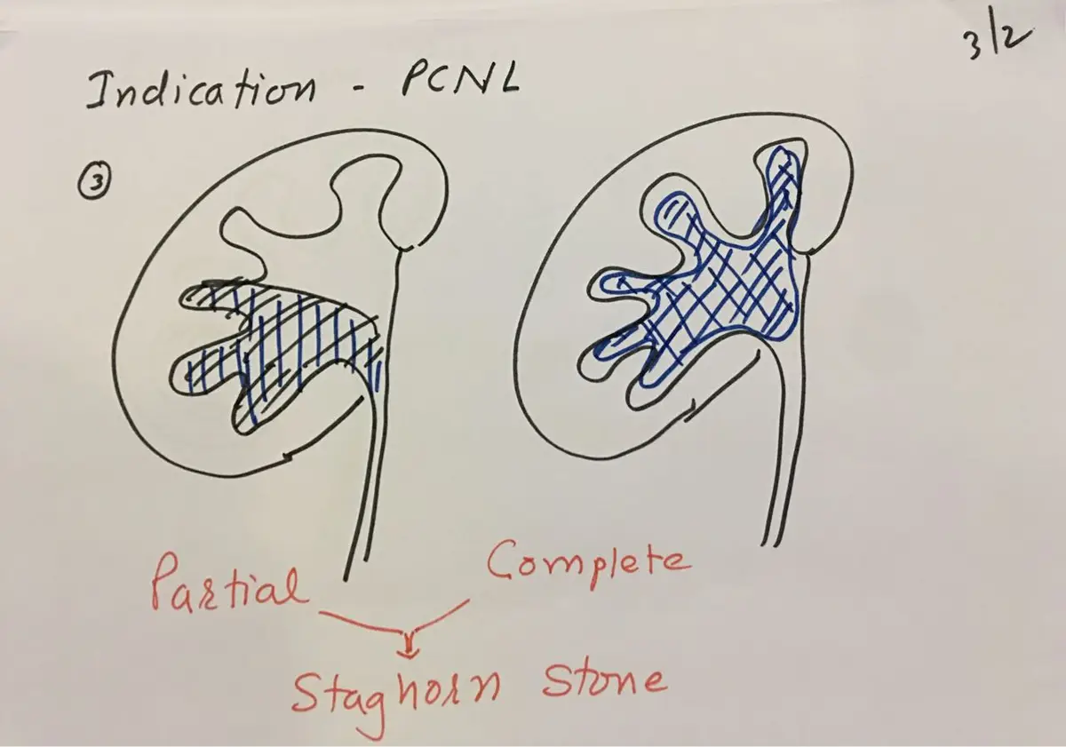
PCNL is the usual choice for kidney stones over 3 cm across, and for staghorn stones, which branch through the kidney's collecting system like antlers, whether complete or partial. It is also used for a retained DJ stent (a soft tube from kidney to bladder) on which a large, complex stone has formed, and for large stones high in the ureter (the tube from kidney to bladder).
Other reasons include a narrowing inside the kidney such as infundibular stenosis (a tight neck to one of the collecting chambers) or a PUJ obstruction (a blockage where the kidney drains into the ureter). PCNL also suits cystine stones over 1.5 cm, stones whose make-up does not respond to shock waves, body shapes unsuitable for shock wave treatment, and stones where shock wave treatment has failed.
When PCNL is not advisable
Some conditions rule PCNL out: a bleeding tendency, pregnancy, an untreated urine infection, a tumour in the path of the planned tract, or a kidney tumour that may be cancerous.
Other situations make it harder and need special planning. These include a horseshoe kidney (two kidneys joined at the lower end), a pelvic kidney (one that sits low in the pelvis), previous open surgery, kyphoscoliosis (a curved spine), obesity, and a closed calyceal neck.
Weighed against these, PCNL is minimally invasive, recovery is short, it is less harmful than open surgery, pain after the operation is usually slight, and the surgeon sees and clears the stone directly. The risks are urine leak, bleeding, stones left behind, the invasiveness of the tract, and a rare chance of needing to remove the kidney.
Planning the way in
Planning the entry is the most important step, both for success and for avoiding complications. I decide the direction of approach to the stone from the layout of the calyces (the cup-shaped chambers that collect urine), how many entries are needed, and the size of the Amplatz sheath (the tube that holds the tract open).
In the face-down position the tract goes into a posterior calyx, one that faces the back. Safety means keeping clear of blood vessels and other organs. For complex stones, a CT scan viewed in three dimensions gives a better plan.
The operation, step by step
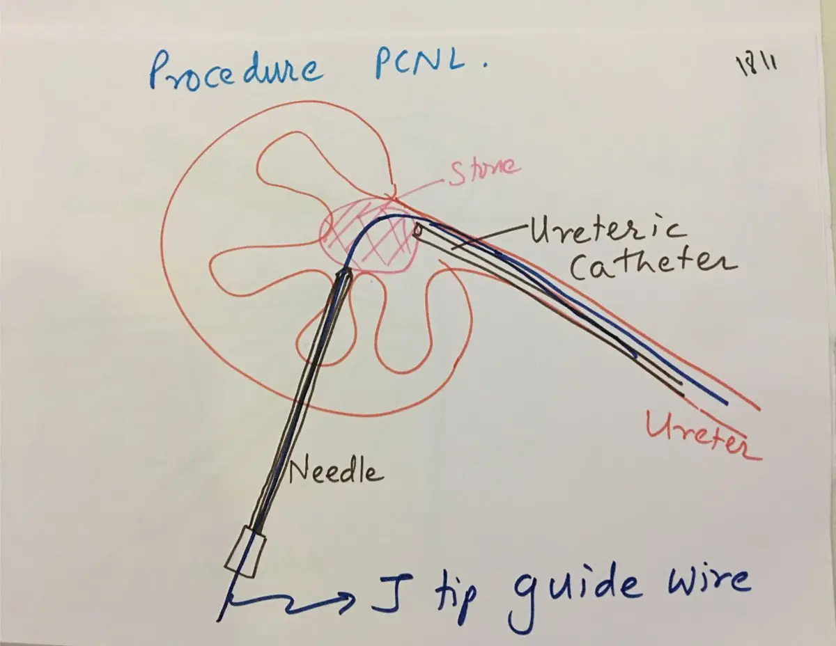
PCNL is done under general anaesthesia, and rarely under spinal or local anaesthesia. First a ureteric catheter, a thin tube, is passed up into the kidney from below. Contrast dye can be injected through it so the collecting system shows up on X-ray. The patient then lies face down or partly on the side.
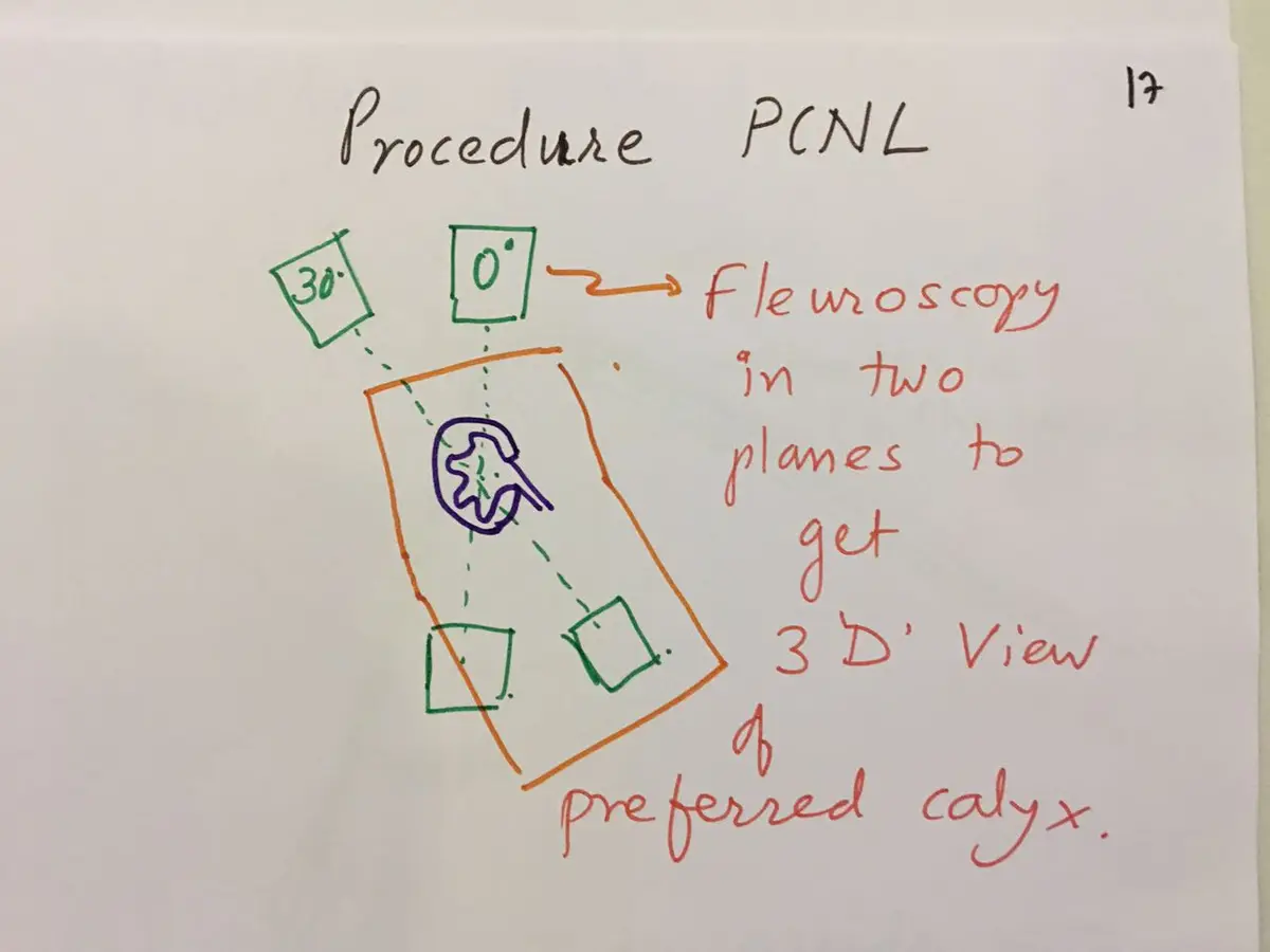
Next I choose the calyx to enter and pass a needle into it, guided by fluoroscopy (live X-ray), ultrasound, or both. With fluoroscopy, taking views straight on and at 30 degrees gives a three-dimensional sense of where the chosen calyx lies.
A J-tip guide wire, with a curved soft end, is threaded through the needle and down into the ureter.
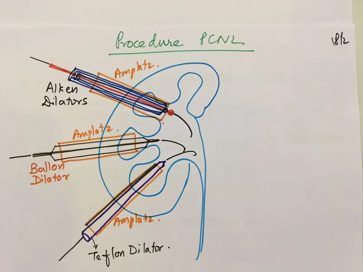
The tract is then widened over the wire with balloon, steel or Teflon dilators, and the Amplatz sheath is slid into place. The optimal sheath size is 22 to 24 Fr (Fr, or French, is a unit of tube width).
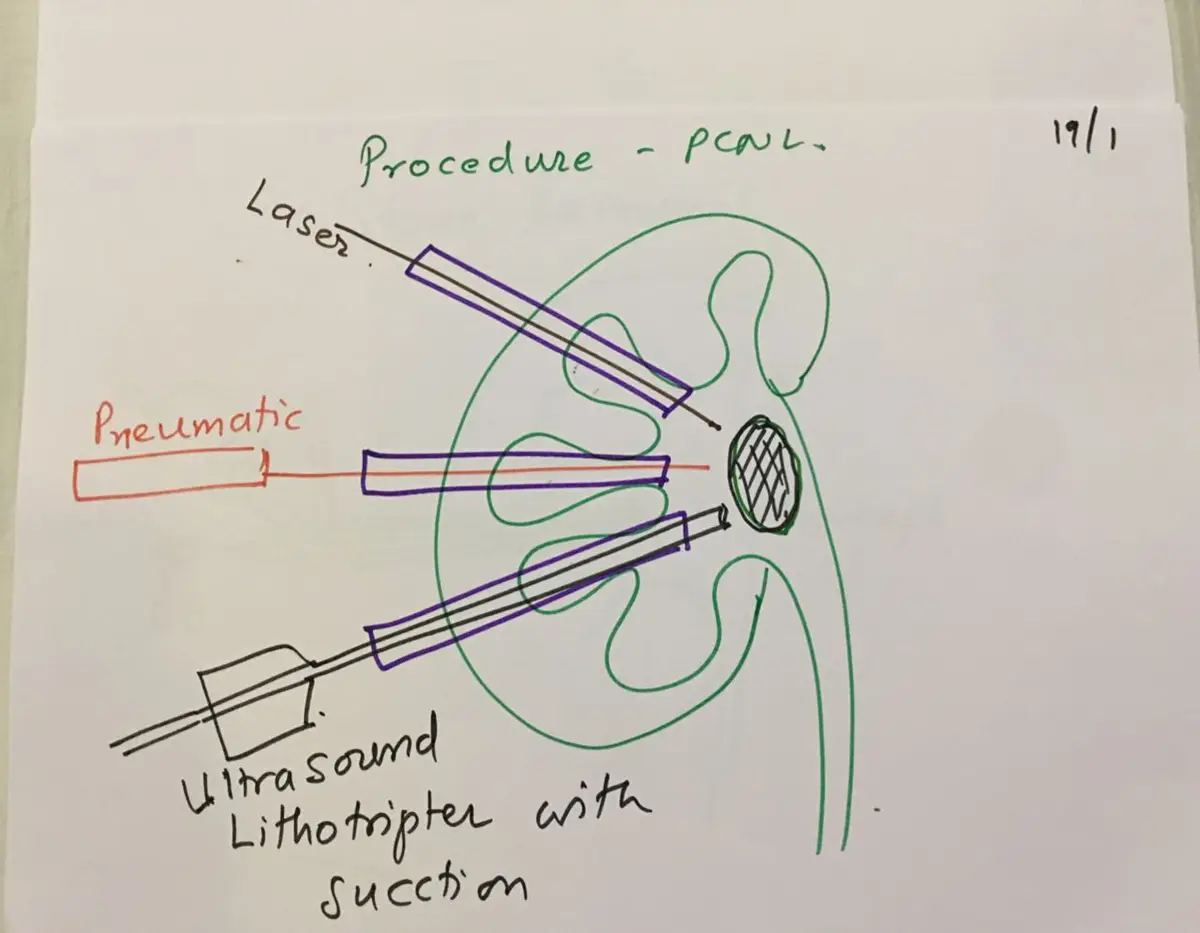
A nephroscope, the telescope for the kidney, goes through the sheath. Scopes come in different sizes and types, rigid and flexible, and the choice is mainly the surgeon's preference. The stone is broken with a pneumatic device, a holmium laser, or an ultrasound lithotripter. The laser gives no kinetic impact, which lessens the chance of bleeding.
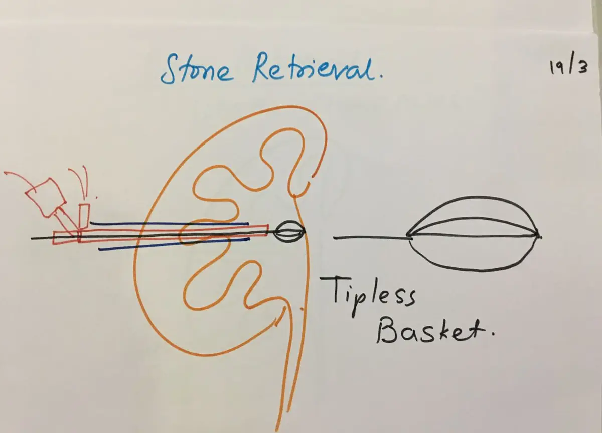
The pieces come out with forceps, with a tipless basket, or by flushing with saline.
Finishing, and dealing with what is left
At the end, the kidney may be drained with a DJ stent, a nephrostomy (a drain from the kidney out through the skin), or nothing at all, which is called tubeless PCNL. The guidelines list factors in the decision, including stones left behind, a likely second look, significant blood loss, urine leakage, blockage of the ureter, infected stones, a single kidney and a bleeding tendency. In uncomplicated cases, going totally tubeless shortens the hospital stay.
A larger residual stone can be reached through another tract. A stone in an adjacent calyx can be reached with a flexible nephroscope or a combined approach from below, and small fragments that cannot be reached can be treated with shock waves or RIRS (a flexible scope passed up from below). If there is bleeding or a perforation, I stop and plan the next sitting after a few days.
The main complications are urine leak, bleeding, fever and residual stone. Bleeding is managed with bed rest, angioembolisation (blocking the bleeding vessel through a catheter), or removal of the kidney.
The talk closed with where PCNL was heading: smaller tracts. Mini PCNL uses a 15/18 Fr system, ultra-mini PCNL 11/13 Fr, and micro PCNL an 18-gauge needle with 8/11 Fr sheaths.