PCNL (percutaneous nephrolithotomy, keyhole surgery through the back or side to remove kidney stones) is the usual treatment for large and complex kidney stones. At IUCC 2026 I taught a workshop on it, titled From Access to Stone-Free Success. Most of it was about the first part of that journey: getting the needle into the kidney safely.
The workshop was the PCNL hands-on training session at the Interdisciplinary Urology Care Consortium (IUCC) conference, held at the Conrad Dubai on 6 and 7 June 2026, where I served as a mentor. The year before, in September 2025, I was a trainer at the PCNL hands-on workshop at ICS-EUS 2025 in Abu Dhabi.
The main steps of every PCNL are the same. We puncture a calyx (one of the cup-shaped chambers inside the kidney that collect urine), widen that puncture into a tract, remove the stone, and plan how to finish safely. This post follows the workshop in that order.
Planning happens before the patient reaches theatre
I ask trainees to imagine the kidney in three dimensions and plan the tract from the CT scan before surgery. The aim is an easy access, a short route through the kidney tissue, and the best chance of clearing all the stone, while avoiding damage to the organs around the kidney.
Planning also means looking at the whole patient. Other illnesses can raise the risk of bleeding and infection, and they affect the choices we make. The size of the tract is planned in advance too.
Three ways to guide the needle
Patients can lie face down (prone) or on their back (supine). The needle can be guided by fluoroscopy (live X-ray imaging), by ultrasound, or by the two together.
With the patient prone, the most common method is the fluoroscopy bull's eye technique. The C-arm, the C-shaped X-ray machine, is used at 0 degrees, then 30 degrees, then 0 degrees again. At 30 degrees the needle is lined up end on over the target calyx, and at 0 degrees its depth is checked. A second method, cranio caudal or triangulation, tilts the C-arm from the head end towards the foot end to judge depth, and it works in both prone and supine positions.
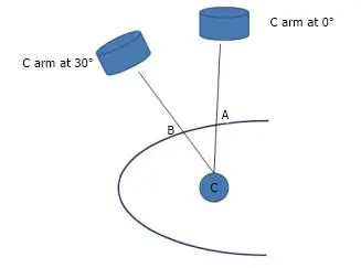
Fluoroscopy is also good at showing when something has gone wrong. If fluid does not come back through the needle, it can be withdrawn gently under X-ray, the puncture restarted, or ultrasound added. Errors such as stopping short of the calyx, passing right through it, or entering the renal pelvis (the central collecting area) or a blood vessel are easy to see on X-ray and may not be clear on ultrasound.
What ultrasound adds
With ultrasound guidance, the kidney and the structures around it are scanned first. The colon and bowel, the liver or spleen, and the pleura (the lining around the lungs) can all be seen and avoided.
The equipment matters. Standard ultrasound probes use an external needle guide set at 35 to 45 degrees. That steep angle makes it hard to line the needle up with the axis of the calyx. It can also leave an air pocket at the edge of the probe and an awkward angle of attack. The newer Fujifilm Arietta 65 machine uses a convex probe with an inline needle guide, which offers puncture angles of 0, 15 and 30 degrees.
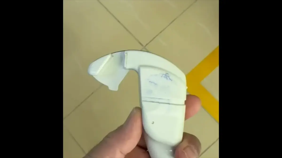
Combining X-ray and ultrasound
In combined guidance, fluoroscopy is kept at a fixed 0 degree angle to show the kidney and the stones, first as a plain image and then with contrast dye. A needle is placed on the patient's skin to mark the line of the puncture. That line then guides the ultrasound: the needle guide mark on the probe is aligned with it.
The echo tip needle, designed to show up clearly on ultrasound, stays visible the whole way in. A final X-ray confirms that the tip has entered the calyx.
Setting up the supine position
For supine PCNL, the patient is first placed in the lithotomy position, lying on the back with the legs raised in supports, so that a ureteric catheter (a thin tube passed up the ureter, the tube that drains the kidney) can be placed. The patient then lies supine, either fully flat or with some upward tilt, so that the needle can enter while avoiding the vital organs around the kidney.
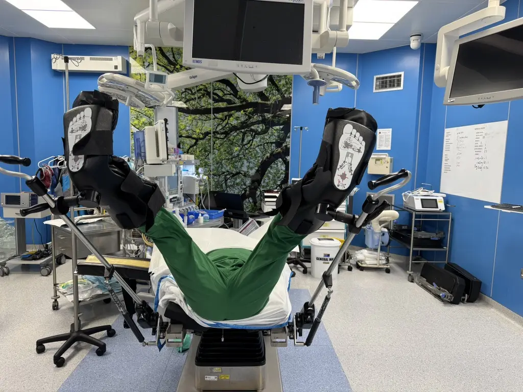
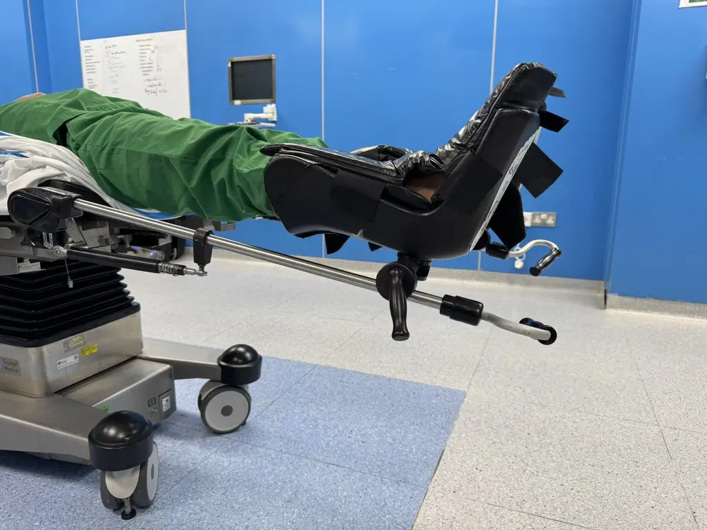
When the anatomy is unusual
Some kidneys need a different plan. A horseshoe kidney, where the two kidneys are joined, is mostly approached prone, and the bull's eye is reversed because the calyces face the 0 degree view. A pelvic kidney, one that sits low in the pelvis, may need laparoscopic guidance, using a camera inside the abdomen. In kyphoscoliosis, a curved spine, a modified position works better, and combined guidance helps.
From the tract to a stone-free kidney
After the puncture, a guidewire is passed into the ureter and the tract is widened with Teflon dilators, Alken metal dilators or a balloon. Dilatation is more accurate under fluoroscopy. A safety wire, placed with the help of the first Alken dilator, is advisable in complex cases, when there is bleeding, and when the tract is twisting or scarred.
Fluoroscopy is then used with the nephroscope (the telescope used inside the kidney) to review stone clearance, and it can guide a flexible nephroscope in a horseshoe kidney. The operation ends with the placement of a stent and a nephrostomy tube.
The hard questions trainees brought
The moments that defined the day were the hard cases, well beyond the textbook ones. What do you do when the wire will not pass into the stone-bearing calyx, the cup-shaped chamber of the kidney that holds the stone? How do you approach PCNL in a horseshoe kidney, where the two kidneys are joined at their lower ends? How do you combine supine PCNL with ultrasound and fluoroscopy fusion in routine practice?
These are the questions that separate a good surgeon from a great one.
Why hands-on training matters
Technique can be taught. Clinical judgment has to be lived. That is why hands-on training matters: it teaches the steps, and it also builds the instincts that kick in when the steps don't work.
To every endourologist in that room: you showed up to get better, and that takes courage. The future of urology is in good hands. I am grateful to have contributed, and grateful to have learned alongside you.
In the workshop I disclosed that I am a key opinion leader for Richard Wolf, EMS Medical Systems and Fujifilm.
What I want trainees to take away is that access is the foundation of the whole operation. A tract that is planned carefully and guided well makes every later step easier, on the way to a stone-free kidney.