Stones can form anywhere in the urinary tract: in the kidney, in the ureter (the tube that carries urine from the kidney to the bladder), in the bladder, or in the urethra. In this talk I covered why they form, how they are found, what people can do to lower their risk, and how we treat them.
Heat, fluids and diet all play a part
Some people are born with a tendency to form stones, and stones can run in families. For most people, though, the causes are closer to daily life: drinking too little, losing fluid through sweating and dehydration, eating a lot of salt, and eating a lot of animal protein, mainly red meat.
Hot, dry weather adds to the risk. So do abdominal obesity and diabetes. Some stones come from metabolic disorders, such as those that produce uric acid or cystine stones, or from renal tubular acidosis (a condition in which the kidneys do not handle acid properly).
How crystals become a stone
Stones are made of crystals. Their main components include calcium oxalate, calcium phosphate, substances linked to bacterial infection, purines such as uric acid, and cystine.
The process starts when urine holds more dissolved salts than it can keep in solution. Doctors call this supersaturation. Tiny crystals then form, clump together, and some are held on the inner lining of the urinary tract, possibly where it has been injured before. The stone keeps growing on these retained crystals. Clumping, or aggregation, appears to be the main way the crystals grow.
Symptoms depend on where the stone sits
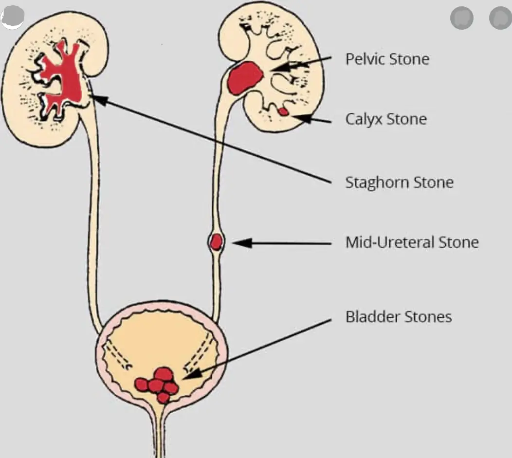
A stone sitting in the kidney may cause no symptoms at all. A stone in the ureter typically causes pain, and may bring red-coloured urine, burning when passing urine, nausea and vomiting. A stone in the bladder or urethra causes difficulty passing urine. If the stone is accompanied by a urine infection, fever can follow.
Finding the stone
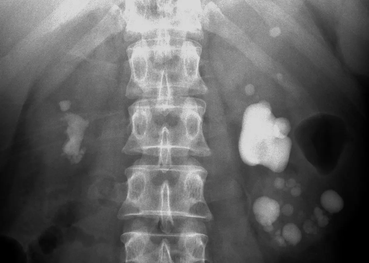
Laboratory tests check how well the kidneys are working and whether there is infection. Imaging shows the stones themselves: an X-ray, an ultrasound, or a CT scan.
Simple habits that lower the risk
Prevention follows directly from the causes. The measures I discussed were drinking enough fluid, avoiding dehydration, and making sure enough urine is passed. On the diet side, they were keeping salt low, keeping red meat and other animal proteins low, and avoiding any type of food in excess. Keeping blood sugar under control and reducing abdominal fat also help.
From medicines to keyhole surgery
Medical treatment starts with managing the pain. Small stones in the ureter can sometimes pass on their own, and a class of medicines called alpha-blockers can help by relaxing and widening the ureter. Medicines that make the urine less acidic, called urinary alkalisers, are used for small stones and for prevention. For uric acid stones, there are medicines that lower uric acid levels.
Whenever possible, we collect the stone and send it for analysis. Knowing what it is made of lets us tailor treatment and prevention to that person.
ESWL (extracorporeal shock wave lithotripsy) uses sound waves from outside the body to break stones in the kidney or ureter.
Endoscopy reaches the stone without any puncture in the body, by passing a thin telescope up through the natural urinary passage. This is called ureteroscopy (URS) when it treats a stone in the ureter, and RIRS (retrograde intrarenal surgery) when a flexible scope goes up into the kidney itself.
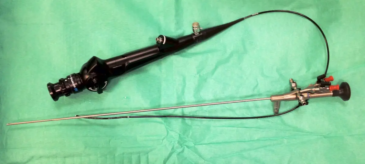
PCNL (percutaneous nephrolithotomy, keyhole surgery through the back to remove kidney stones) reaches the stone by directly puncturing the kidney through a small opening in the skin.
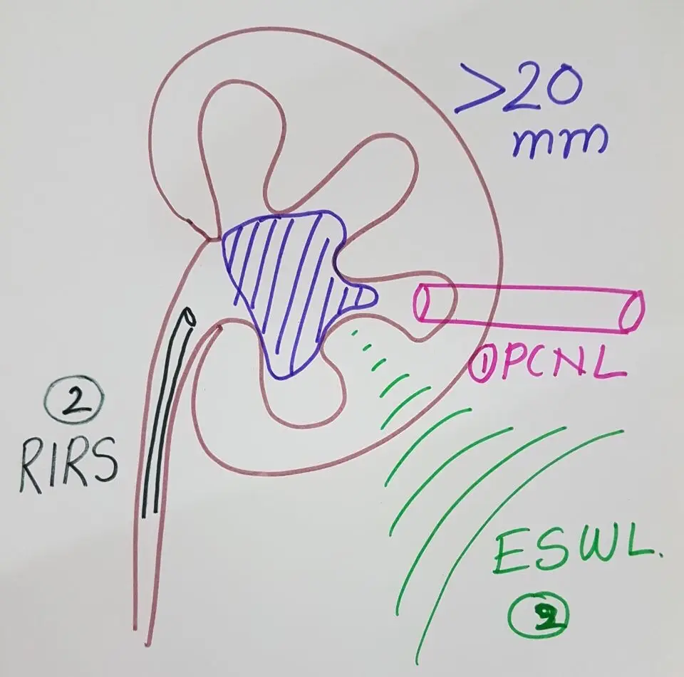
The choice between these options depends on the size and position of the stone and on the person being treated, and it is a decision the urologist makes together with the patient.
If you have pain in your side or back, blood in the urine, or difficulty passing urine, please see a urologist.