PCNL (percutaneous nephrolithotomy, keyhole surgery through the back or side to remove kidney stones) depends heavily on its equipment. Each stage of the operation has its own set of tools, and knowing what each one does, and when to reach for it, matters most with complex stones.
In November 2018 I gave the instruments lecture in the PCNL instruction course at the 7th Emirates International Urological Conference. My aim was to go through the instruments used in the technique and discuss how to use them for complex stones. This post follows the same order as the operation.
Setting up the room
Before any instrument touches the patient, the room has to be right. For PCNL with the patient lying on their back (the supine approach), we need a radiolucent table, one that X-rays pass through so the kidney can be imaged during surgery. Pressure points are protected with proper supports and cushions, and the anaesthetic breathing tube and intravenous line need care while the patient is positioned. PCNL can also be done with the patient lying face down, called prone PCNL.
The surgeon works with monitors for the endoscope camera, the ultrasound and the fluoroscopy (live X-ray) all in view. The instrument trolley is prepared with everything the case will need.
The first step starts from below
The operation begins with cystoscopy, where a telescope is passed into the bladder, followed by ureteric catheterisation. A thin ureteric catheter is passed up the ureter (the tube draining the kidney into the bladder) towards the kidney.
Finding the right calyx
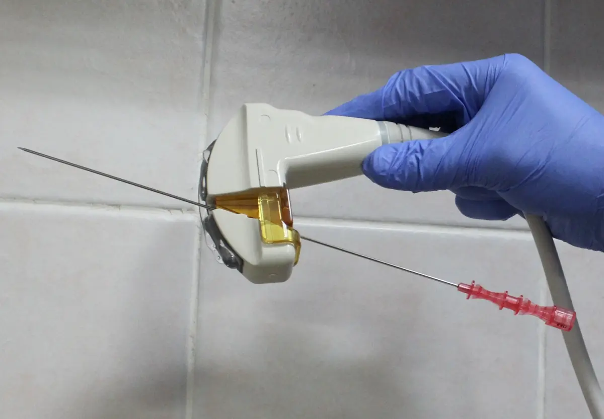
Next comes the puncture. The surgeon identifies the preferred calyx (one of the cup-shaped chambers inside the kidney that collect urine) and passes a needle into it through the skin. The needle I showed is a two-part, 18 gauge puncture needle.
Guidance for the puncture can be by fluoroscopy, by ultrasound, or by the two combined. For ultrasound, a needle guide attaches to the probe and the needle passes through it. On fluoroscopy, the X-ray arm is used both straight on (0 degrees) and tilted to 30 degrees.
Widening the path into the kidney
Once the needle is inside the collecting system, a J-tip guide wire (a flexible wire with a curled end) is passed through it and down into the ureter.
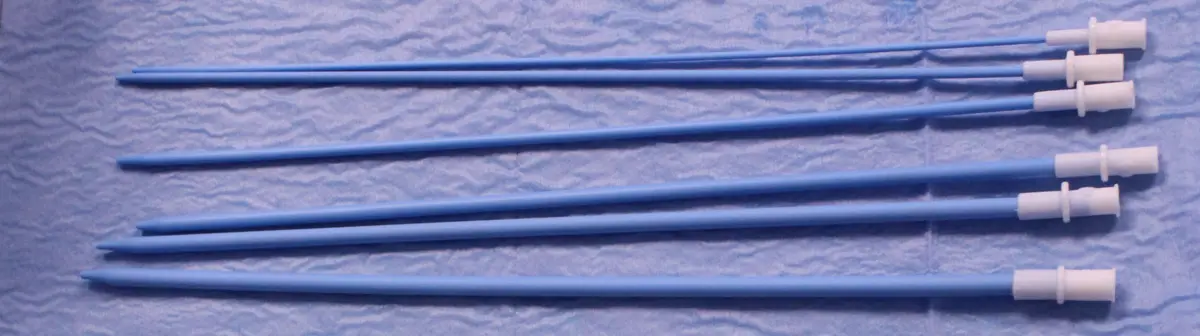
The tract (the passage from skin to kidney) is then widened in steps. The options are balloon dilatation, steel dilators, or Teflon dilators. The Teflon set runs from 6 to 16 French. French (Fr) is the scale used for the width of these instruments. The steel Alken dilators pass over a guide rod.
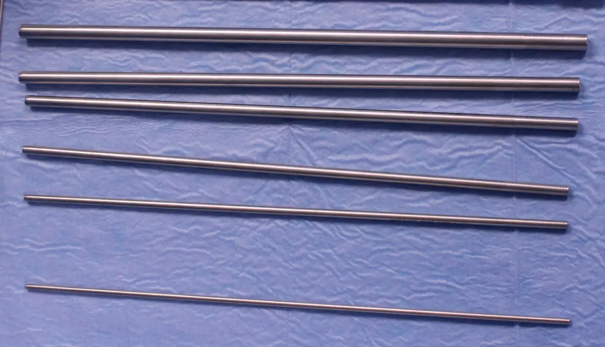
At the end of dilatation, an Amplatz sheath (a rigid working tube that holds the tract open) is placed. The optimal size is 22 to 24 Fr.
Choosing the telescope
The nephroscope is the telescope used inside the kidney. Its size and type determine how the stone is seen, how it is broken, and how the pieces are brought out. Flexible endoscopes are part of that choice too.
There are four broad groups. The standard nephroscope is larger than 19 Fr. Mini PCNL uses 15/18 Fr. Ultra-mini PCNL (UMPL) uses 11/13 Fr. The Micro Perc system (needle PCNL) works through an 18 gauge needle, with 8 or 11 Fr options.
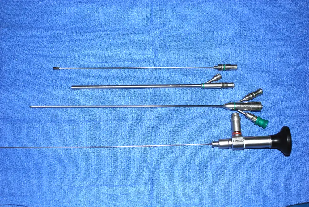
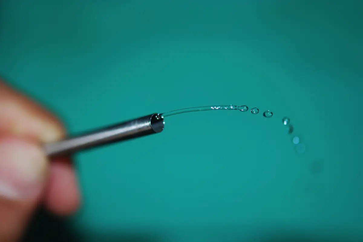
In the smaller systems, stone retrieval works differently. Saline is pushed through a small inner tube as a jet, which creates a whirlpool effect, and the fragments are flushed out through the outer sheath. In the Micro Perc system the needle sheath carries the scope, the laser fibre and the saline together.
Breaking and removing the stone
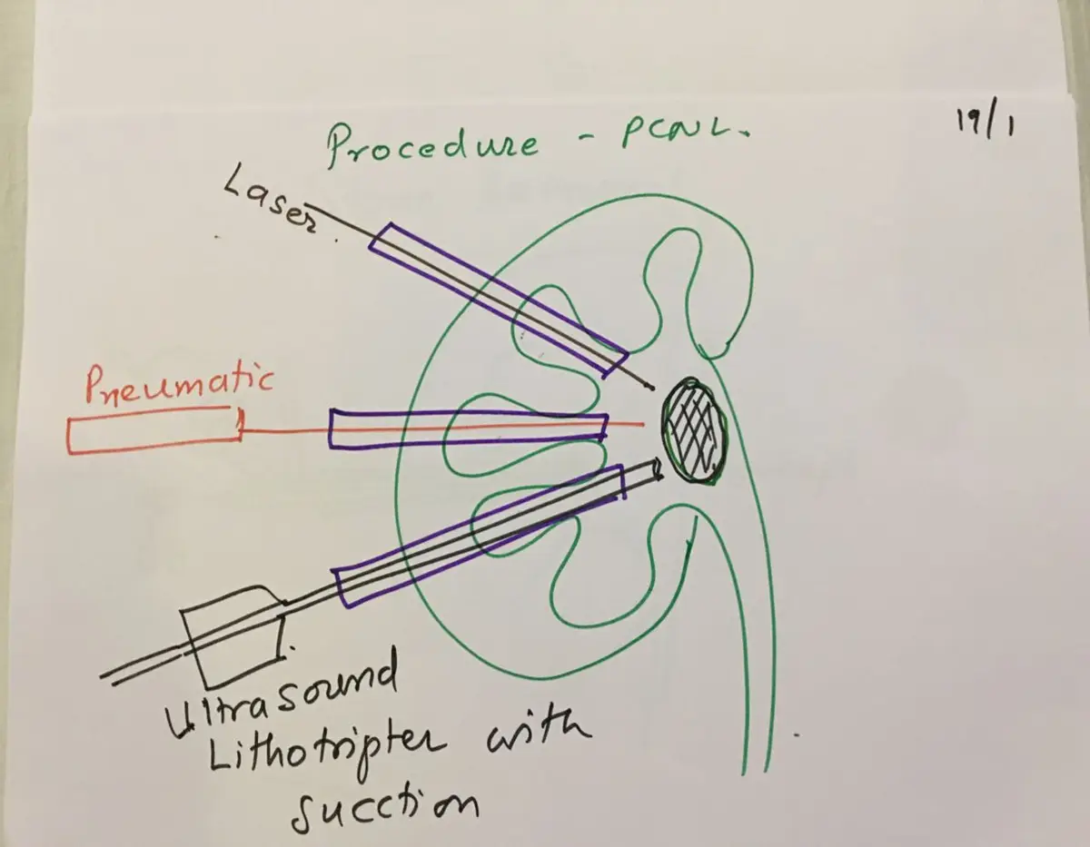
Once the stone is in view, it has to be broken. The energy sources I covered were the laser, the pneumatic lithotripter (which breaks the stone by mechanical impact, like a small jackhammer), and the ultrasound lithotripter with suction, which breaks and removes fragments at the same time.
Forceps grasp and remove the larger fragments. A flexible nephroscope can bend into calyces the rigid scope cannot reach, and in a combined approach a second scope comes up from below through the ureter. At the end, a nephrostomy tube (a drain from the kidney out through the skin) may be placed.