Large and complex kidney stones are usually treated with PCNL (percutaneous nephrolithotomy, keyhole surgery through the back to remove kidney stones). At the 5th Interdisciplinary Urology Care Consortium in Dubai, I took part in a debate on how that surgery should be guided: with fluoroscopy (live X-ray imaging in the operating theatre) or without it, relying on other imaging such as ultrasound.
My position was the case for fluoroscopy. This post sets out that argument, and also the steps I take to keep radiation to a minimum, because any honest case for X-ray guidance has to deal with its main drawback.
Fluoroscopy has guided PCNL from the start
Every PCNL follows the same main steps: puncturing the right calyx (one of the cup-shaped chambers that collect urine inside the kidney), widening that puncture into a tract, dealing with the stone, and planning the exit at the end. Fluoroscopy has been part of PCNL since the operation was invented.
The history explains why. After the Second World War, image intensifiers and the C-arm (the C-shaped X-ray machine used in theatre) made images around 500 times brighter. In 1976 Fernström and Johansson removed kidney stones through a nephrostomy (a tube placed through the skin into the kidney) under radiological control. The first CT scanner arrived in 1971, and CT later improved planning before surgery and checking for leftover stones afterwards.
Anatomy mattered as much as imaging. In 1990 Francisco Sampaio studied casts of the kidney's drainage system and its blood vessels. His work supports entering the kidney through the fornix, the outer rim of a calyx, and avoiding the infundibulum, the narrow neck of the calyx, to minimise the risk of bleeding. So before any puncture, I review the CT scan and plan the route.
A good operation starts with a good puncture
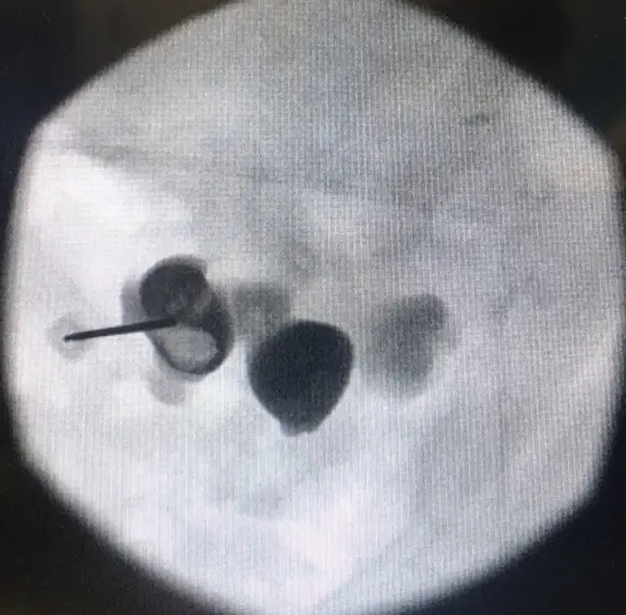
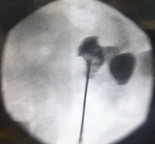
There are three ways to guide the needle: fluoroscopy alone, fluoroscopy combined with ultrasound, or ultrasound alone. With the patient lying face down, the most common fluoroscopy method is the bull's eye technique. The C-arm is used at 0 degrees, then 30 degrees, then 0 degrees again.
At 0 degrees I check the line of the puncture. At 30 degrees the needle is lined up end on, like looking down a barrel, so that its tip and hub sit over the same point on the target calyx. Back at 0 degrees I confirm the final depth of entry. The other approach is the cranio caudal or triangulation technique, where the C-arm is tilted from head to foot to judge depth. I use it both face down and with the patient lying on their back (supine).
Where X-ray guidance solves real problems
Fluoroscopy earns its place when things are difficult. If two calyces sit close together, I can try both the 0 and 30 degree method and the cranio caudal method. If no fluid comes back through the needle, I can withdraw it gently under X-ray, start again, or bring in ultrasound as well.
Errors in the puncture are also easy to see on fluoroscopy, and may not be clear on ultrasound. These include stopping short of the fornix, passing right through the calyx, ending up in kidney tissue between two calyces, entering the renal pelvis (the central collecting area) directly, or entering the renal artery or vein.
Beyond the puncture: dilatation, clearance and the exit
After the puncture, a guidewire is passed down into the ureter (the tube that drains the kidney to the bladder) and the tract is widened. Depending on the case, I use Teflon dilators, Alken metal dilators or a balloon. The first Alken dilator can also be used to place a second, safety guidewire, which I advise in complex cases, when there is bleeding, and when the tract is twisting or scarred. Dilatation is more accurate when it is watched on X-ray.
During stone removal, fluoroscopy combined with the nephroscope (the telescope used inside the kidney) helps me review whether stones are cleared. In a horseshoe kidney, where the two kidneys are joined at the lower end, it helps guide a flexible nephroscope through the working sheath.
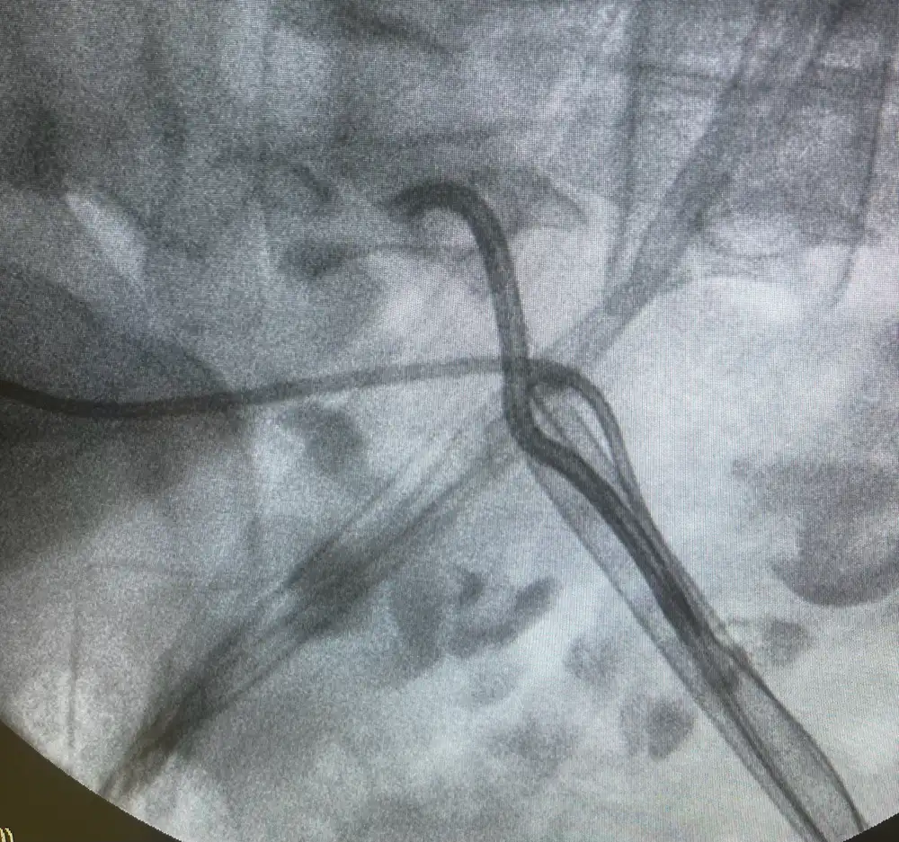
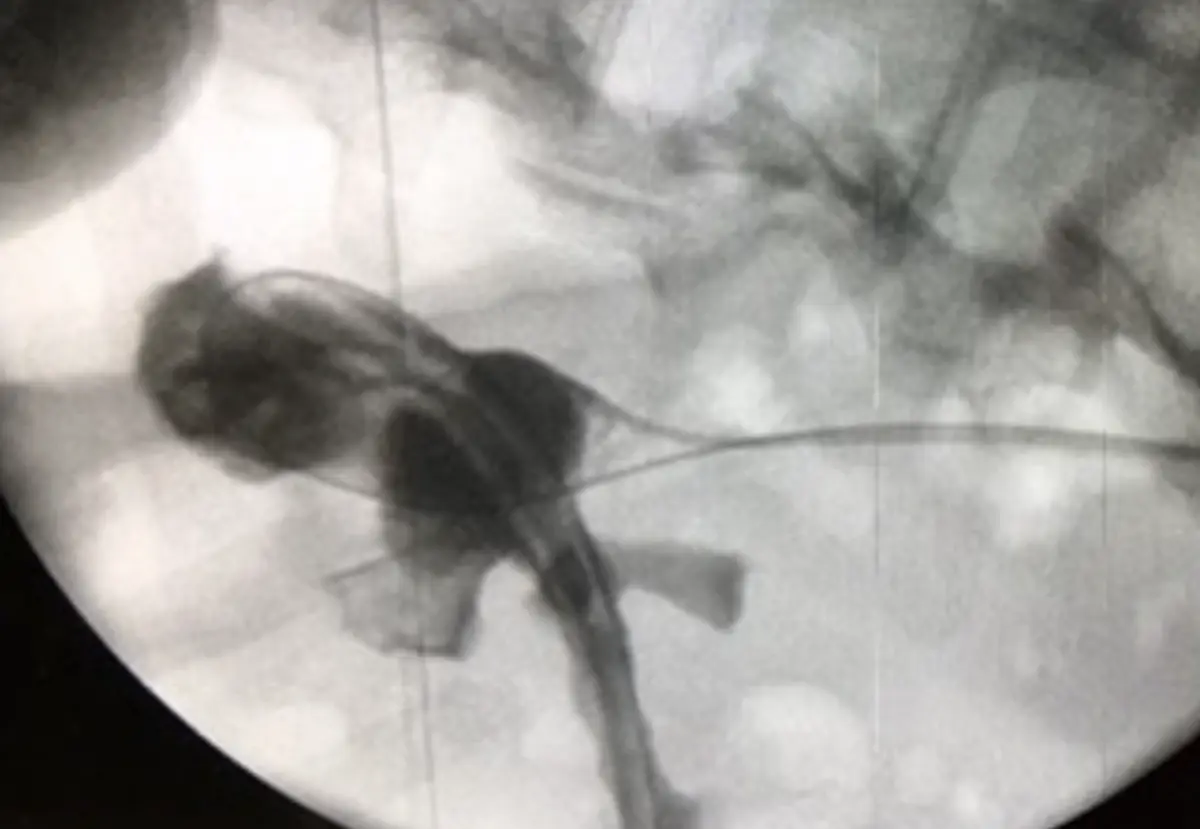
At the end, it confirms the position of the drainage tubes: a DJ stent (a thin tube running from the kidney to the bladder) and a nephrostomy tube held with the balloon of a Foley catheter. A final nephrostogram, an X-ray taken with contrast dye, shows how things look before we finish.
Keeping the radiation dose low
Radiation is the fair objection to fluoroscopy. Its hazards include cancer, heritable effects in offspring, and injury to the skin, bone and eye. The team is exposed to both direct and scattered radiation. Studies of operator exposure show that when the imaging field sits near the patient, exposure is high, and when it sits further away, the operator's upper body receives less.
Set-up makes a difference. A table set too low increases the dose entering the patient and the scattered dose reaching the operator. A table too low with the detector too high increases the patient's dose further. The imaging mode matters too: standard fluoroscopy uses around 20 to 40 nGy per frame, cine acquisition around 200, and digital subtraction around 1,200.
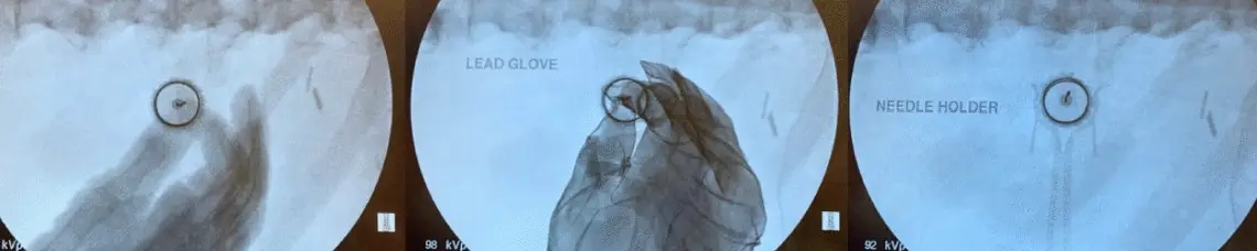
The measures I follow are simple. Use ultrasound where it can do the job. Keep the beam on for as short a time as possible and use the lowest dose, slowest frame rate setting. Narrow the X-ray beam to the area of interest, keep the detector close to the patient, and use as much shielding as possible. I also keep my hands out of the beam by holding the needle with a metal grasper.
Why fluoroscopy still belongs in the theatre
My argument in the debate was that fluoroscopy supports every stage of PCNL, from the first puncture to the final check, and makes errors visible as they happen. Ultrasound has a real role, alone or combined with X-ray, and it lowers the radiation dose. Used with care, fluoroscopy remains a dependable guide for safe and accurate keyhole kidney stone surgery.