Endourology is surgery done through the body's own passages or through small keyhole tracts, using telescopes instead of open incisions. Urolithiasis is the medical name for stones in the urinary tract. This talk covered how the endourological treatment of kidney stones has changed, and how I choose between the options.
Each new technique has pushed the last one aside
In the late 1970s surgeons began using a tract through the skin to remove kidney stones, and Arthur Smith pioneered PCNL (percutaneous nephrolithotomy, keyhole surgery through the back). Then ESWL (shock waves from outside the body to break stones) took over from PCNL for many stones. RIRS (a flexible telescope passed up through the urinary passage into the kidney) in turn took ground from ESWL. Most recently, miniaturised PCNL has taken ground from ESWL.
Today the options are ESWL, PCNL, RIRS, a combined approach using PCNL and RIRS together, and laparoscopy. With so many choices, the real question is which one suits a particular patient and stone.
Stone size and position guide the first choice
Before planning, I look at the patient's other illnesses, the site, size and shape of the stone, and the anatomy of the kidney. Stones need active removal when they grow, block the kidney, cause infection or symptoms such as pain or bleeding, or occur in patients at high risk of forming more stones.
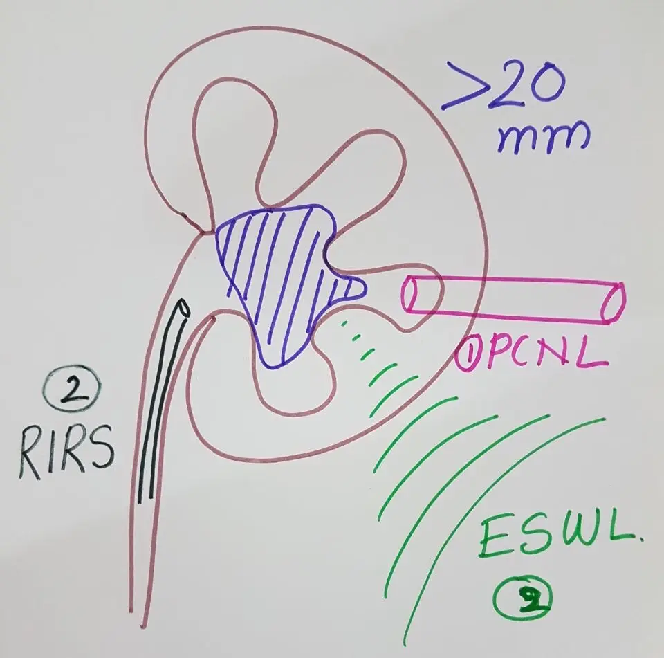
For a kidney stone over 20 mm, the guideline options are PCNL, RIRS, a combined approach, or ESWL with a DJ stent (a soft tube from kidney to bladder); laparoscopy and open surgery remain in reserve. For stones of 10 to 20 mm outside the lower part of the kidney, ESWL or endourology, meaning mini PCNL or RIRS, are the choices.
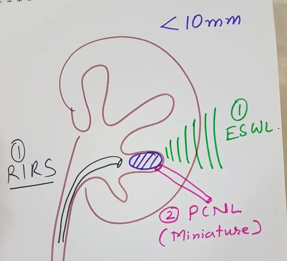
For stones under 10 mm, ESWL or RIRS come first, with the smallest forms of PCNL as the second line. Stones of 10 to 20 mm in the lower calyx (the lowest collecting chamber of the kidney) depend on whether conditions favour ESWL. If they do, ESWL comes first; if they do not, endourology comes first.
Small stones of about 5 mm that cause no symptoms and are stable are managed without surgery, with medicines, diet and regular ultrasound follow-up.
Standard PCNL, and the move to smaller tracts
PCNL is still the standard procedure for large kidney stones. It is minimally invasive, recovery is short, it is less harmful than open surgery, and it lets the surgeon see and clear the stone directly. Its risks include urine leak, bleeding, infection and injury to other organs, with a rare chance of losing the kidney.
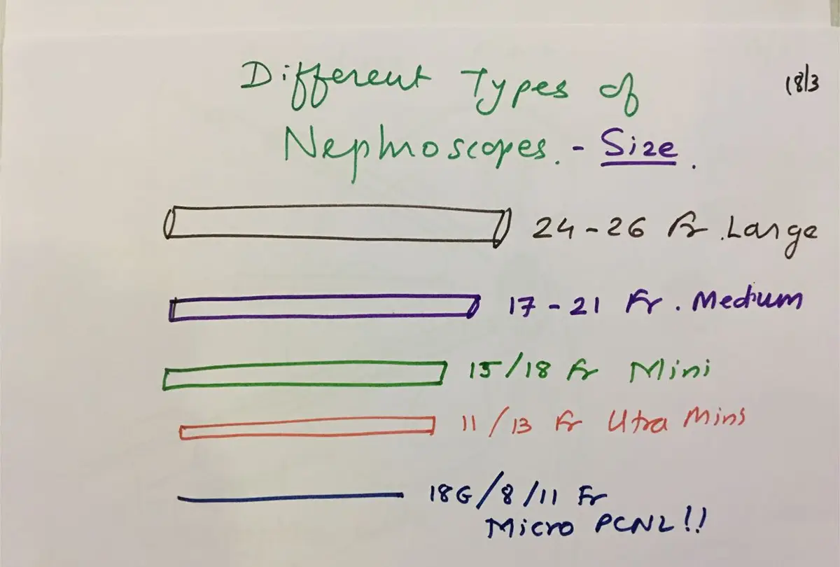
The tract size has shrunk over time. Early PCNL used a 32 Fr sheath (Fr, or French, is a unit of tube width). Standard PCNL now uses a 22 or 24 Fr Amplatz sheath, the tube that holds the tract open. Standard PCNL suits larger stones, allows several punctures for staghorn stones (stones that fill the kidney's branches), and takes less time to clear stone. Its downsides are a possibly higher chance of bleeding and more pain after surgery.
Smaller tracts bring their own trade-offs: longer procedures and poorer vision because less saline flows through, with possibly less bleeding and pain. The guidelines at the time noted that miniaturised systems seemed effective, but that their benefit over standard PCNL was still to be proven and smaller instruments can raise pressure inside the kidney.
Mini, ultra-mini and micro PCNL
Mini PCNL uses a 15/18 Fr system. It is an established technique, and many centres now do only mini PCNL. Access is faster, with less pain and possibly less bleeding. The instruments and forceps are smaller, stones are flushed out with saline, and stone clearance may take longer.
Ultra-mini PCNL (UMPL, 11/13 Fr) and micro PCNL are for stones up to 18 mm in the lower calyces, in calyces with a narrow neck, stones RIRS cannot reach, and fragments left after PCNL that the telescope cannot reach. As with every PCNL, the puncture is the most important step: if the needle misses the calyx, the tract may not reach the stone.
The UMPL set I showed is autoclavable (it can be steam sterilised), with a 3.5 Fr, 1 mm scope of 17,000 pixels. A holmium laser breaks the stone, and fragments are flushed out through the outer sheath.
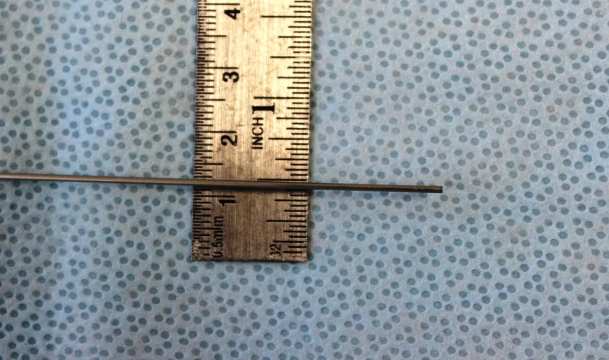
Micro PCNL, also called needle PCNL, goes further. The scope is 0.9 mm with 10,000 pixels and passes through a disposable needle, and the laser fibre is up to 350 microns. The advantages I listed were fast access, no tube, no pain and no bleeding. The limitations are that no stone is retrieved for analysis, fragments have to pass on their own, conversion to a larger sheath may be needed, and the disposable cost is higher than UMPL.
Finishing the job: residual stones, tubes and combined surgery
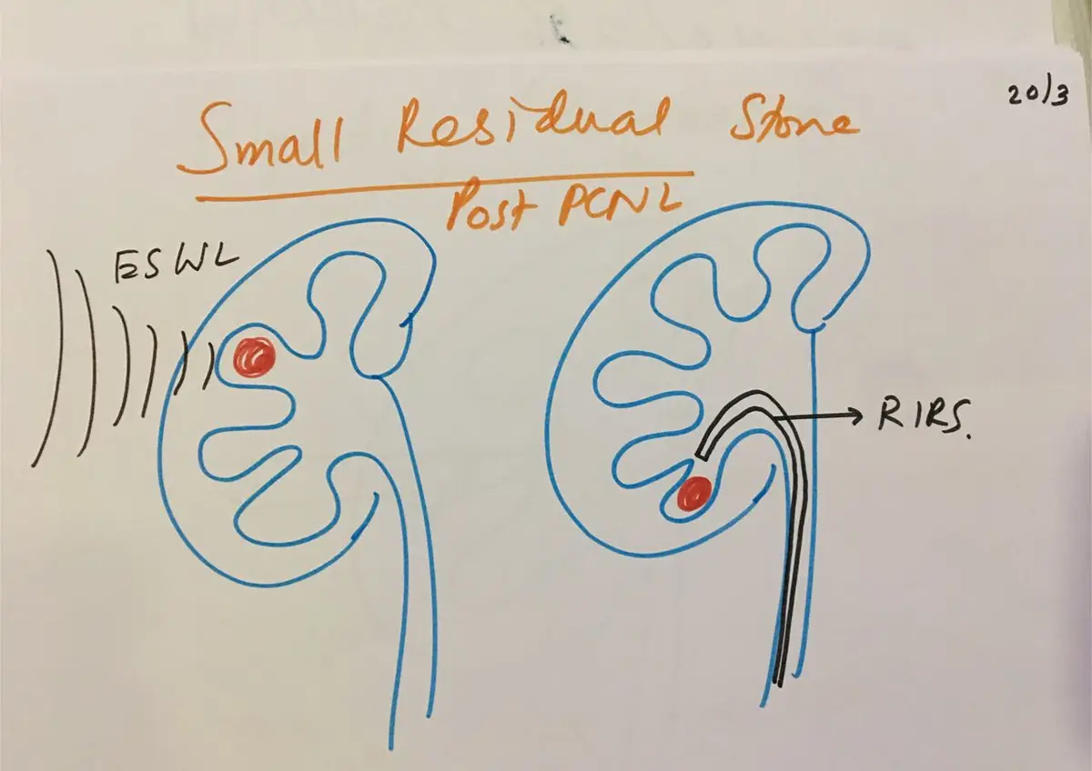
A larger stone left behind can be reached through another tract. A stone in an adjacent calyx can be reached with a flexible nephroscope or a combined approach, and small inaccessible fragments with ESWL or RIRS. If there is bleeding or a perforation, I stop and plan a second sitting a few days later.
At the end of PCNL, the kidney can be drained with a DJ stent, a nephrostomy tube (a drain from the kidney through the skin), or neither. Tubeless PCNL leaves no nephrostomy; when neither a nephrostomy nor a stent is used, it is called totally tubeless. The guidelines noted that in uncomplicated cases this shortens the hospital stay.
For scattered stones with a large total burden, I combine the approaches. I start with a telescope from below to break the stones and move them to the renal pelvis or one calyx, while the kidney stays filled with saline. Then I do a supine PCNL (with the patient lying on their back) to remove the fragments. This avoids multiple punctures, and stones come out easily with gravity, though it needs two teams and two sets of equipment.
The conclusion of the talk was simple. No one procedure is the gold standard for every stone. Each patient needs a tailor-made plan, decided together, considering all the factors and the patient's wishes.